Our quantum materials research laboratories are equipped with state-of-the-art equipment for materials synthesis, structural characterization, electronic structure studies, and physical property measurements. These facilities are all located at the IAMM headquarters. Complementary research capabilities are offered through UT’s high-performance computing infrastructure, as well as ORNL’s supercomputing center and neutron sources such as HFIR and SNS.
Synthesis
Nearly all discoveries in materials research start with the synthesis of novel compounds. UT-IAMM houses one of the largest solid-state synthesis laboratories in the nation with a wide variety of furnaces for the synthesis of polycrystalline materials and large single crystals. Our motto: those who control the synthesis direct the future of materials science.
Principal Investigators: David Mandrus, Yishu Wang, Haidong Zhou
Epitaxial Synthesis
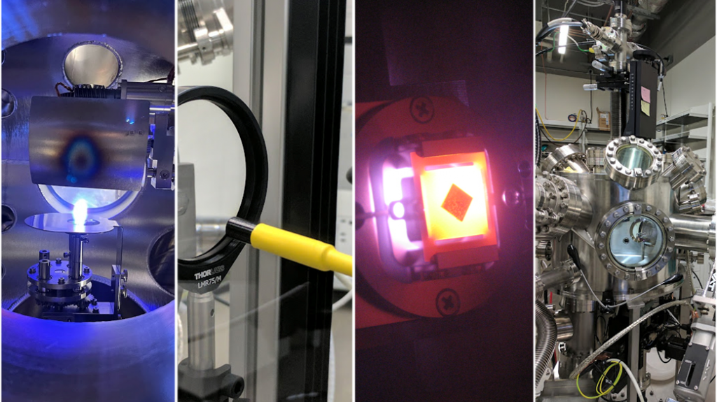
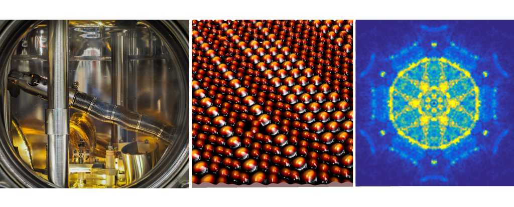
Molecular beam epitaxy is the most advanced thin film synthesis technique for the growth of single-crystalline thin films with the highest purity and atomically precise control of the thickness and composition. The quantum materials group employs MBE synthesis of transition metal oxides and chalcogenides as well as conventional group III-V semiconductors. It also employs pulsed laser deposition (or ‘laser MBE’) of complex oxide materials that are very difficult to grow via thermal MBE. The oxide MBE apparatus is an IAMM core facility.
Principal Investigators: Profs. Joon Sue Lee, Jian Liu, and Hanno Weitering
Electron Spectroscopy
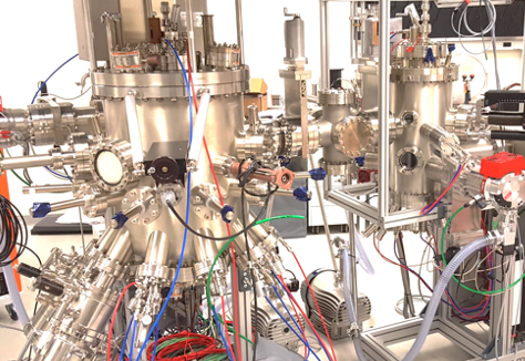
Dual anode (Al/Ag) XPS apparatus with x-ray monochromator and spin detector. This particular instrument is an IAMM core facility.
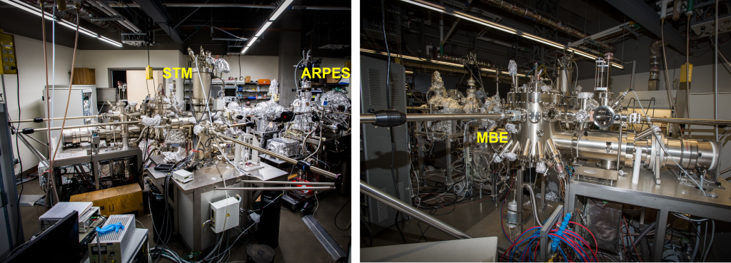
The electronic properties of quantum materials are studied with various electron spectroscopies such as x-ray photoelectron spectroscopy (XPS), Auger electron spectroscopy, angle-resolved photoemission (ARPES) with time-resolution capability, and high-resolution electron energy loss spectroscopy (HREELS) in reflection mode. These techniques provide direct information on the electronic structure and excitations in quantum materials. The photoelectron spectrometers are part of an ultrahigh vacuum suite with added capabilities for molecular beam epitaxy and scanning tunneling microscopy.
Principal Investigators: Prof. Norman Mannella and Hanno Weitering
Topographic and Spectroscopic Surface Imaging
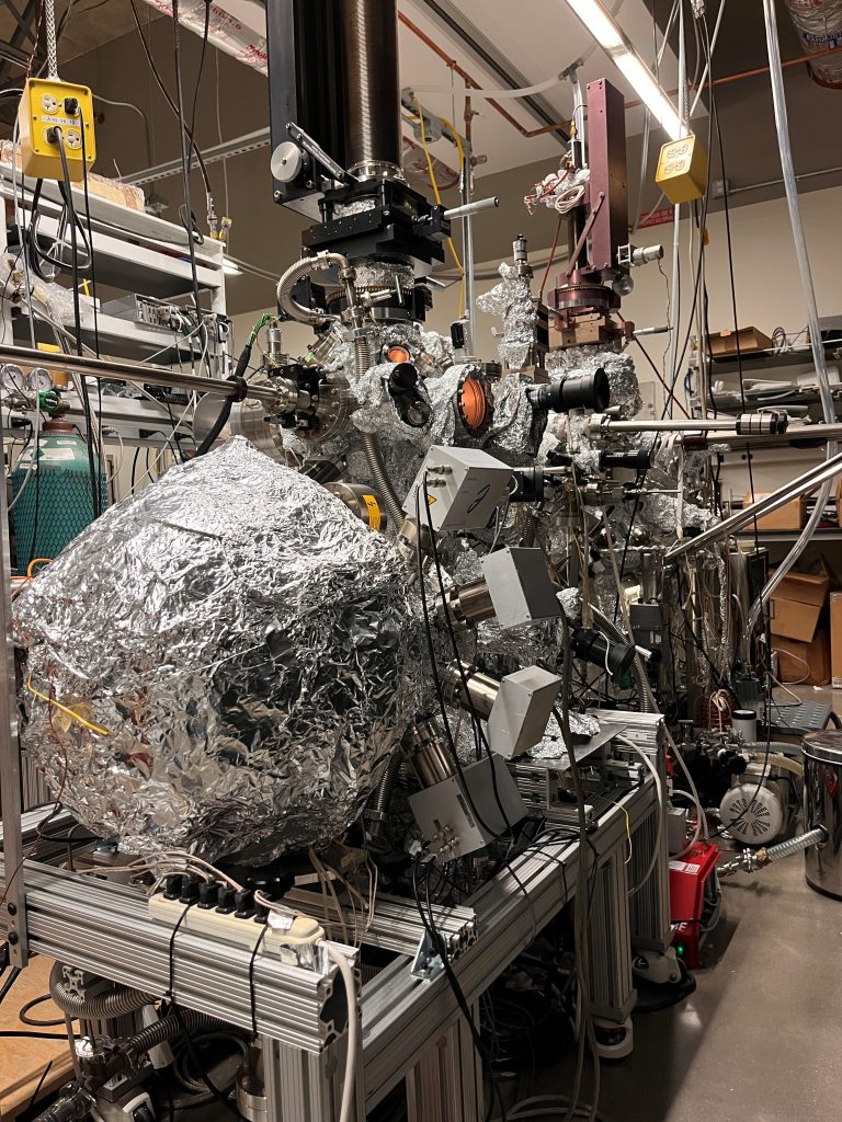
Scanning Tunneling Microscopy is a very powerful tool for studying the topography of a crystal surface or thin film with atomic resolution. In topographic imaging, one applies a small bias and measures a small quantum mechanical tunneling current between an atomically sharp metal tip and the crystal surface as a function of tip location above the surface. The latter can be controlled with Angstrom precision. It also is a spectroscopic tool that can measure the local electronic density of states (proportional to the dI/dV signal) as a function of applied bias and tip location. Differential conductance maps provide key information about, e.g., the Fermi surface of the material, presence of topological edge states, or symmetry of the order parameter in superconductors.
Principal Investigators: Profs. Wonhee Ko and Hanno Weitering
Contact: Dr. Paolo Vilmercati
Electron Microscopy
IAMM’s Microscopy Core Facility houses a variety of scanning probe and electron microscopes, including an atomic force microscope (AFM) and a Nano-indenter, two transmission electron microscopes, a scanning electron microscope and two Dual Beam Focused Ion Beam (FIB) instruments. One showcase instrument is the 300 keV ThermoFischer monochromated and aberration corrected TEM/STEM with spatial resolution down to 40 pm, allowing atomic resolution EDS and EELS. Another showpiece is a Plasma FIB providing ion beams of Xe, Ar, N, and O, for imaging and processing of prototypical devices. This instrument is equipped with EDS and EBSD for microstructural and chemical analysis.
ThermoFischer Spectra 300 aberration corrected TEM/STEM at IAMM’s Microscopy Facility.
Structural Characterization
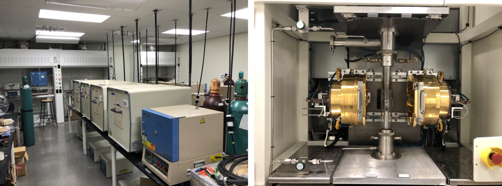
Characterization of new materials usually starts with structure determination via x-ray diffraction. IAMM’s Diffraction core facility houses a variety of x-ray diffractometers. Basic usage of these machines revolves around the identification and quantification of crystalline phases, space group symmetry and lattice parameters, crystallite size, and micro strain. More advanced uses include high-temperature in-situ experiments, investigating preferred orientation with pole figures, determining residual stresses, and studying thin films with X-ray reflectivity, among others.
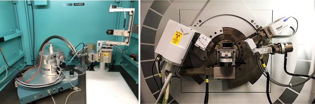
Left: X-ray machine for powder diffraction and Laue back reflection for crystallographic orientation. Powder diffraction data can be recorded at temperatures as low as 10 Kelvin. Right: Empyrean diffractometer from Malvern Panalytical, used for routine powder diffraction experiments. Special features include the availability of three different wavelengths of radiation (copper, cobalt, and silver) and the ability to perform in situ high-temperature diffraction experiments up to 1200 Celsius in air, inert gas, and vacuum environments. The Empyrean was recently upgraded to allow for pair distribution function measurements.
Principal Investigators: David Mandrus, Yishu Wang, Haidong Zhou
Electrical and Magnetic Property Measurement
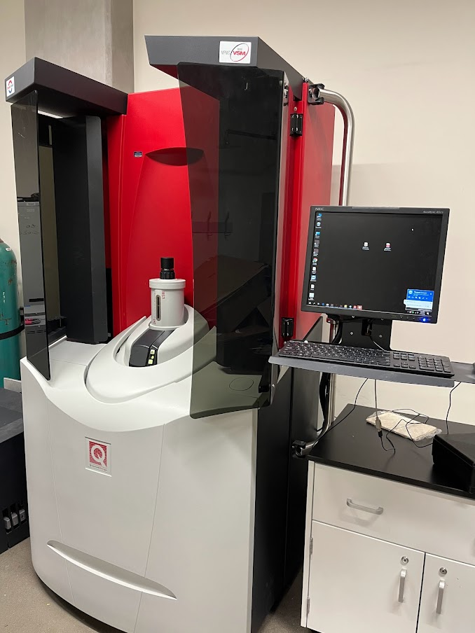
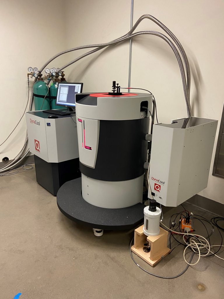
Cryo equipment for measurements of electrical, magnetic and other physical properties
Quantum materials exhibit a wealth of fascinating electrical and magnetic properties. These can be measured using our Quantum Design Physical Properties Measurement Systems (PPMS) and MPMS-VSM magnetometer system. We routinely access temperatures as low as 2 Kelvin and magnetic fields up to 14 Tesla. The magnetometer is equipped with a Helium-3 option for magnetization measurements down to 0.4 Kelvin. These instruments are part of IAMM’s core facilities.
Principal Investigators: David Mandrus, Jian Liu, Yishu Wang, Haidong Zhou
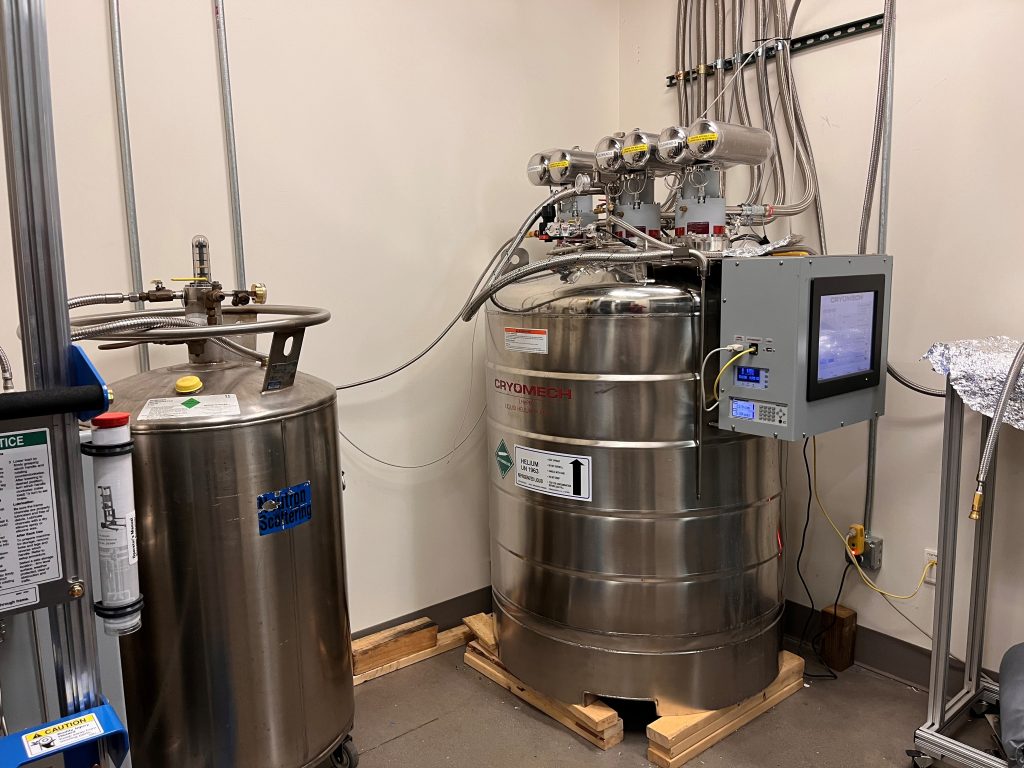
Research at low temperatures
Many experiments are done at low temperatures and require liquid helium cooling, which is an increasingly rare and expensive commodity. To reduce the cost of low-temperature experiments, our researchers have access to a central helium recovery and liquefaction facility capable of producing 60 liters of liquid helium per day. The system is equipped with a 500-liter storage dewar.
Neutron Scattering
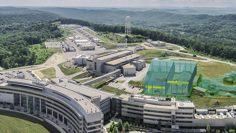
Neutron scattering is a powerful way to probe the magnetism and therefore quantum behavior in materials. We principally use the Spallation Neutron Source and High Flux Isotope Reactor, two of the World’s premier neutron facilities located at Oak Ridge National Laboratory for our experiments. We both undertake experiments on the structure and dynamics of materials as well as push scientific boundaries by developing new techniques and sample environments. Much of our work utilizes the Shull Wollan Center, the UT Center for neutron science on the Oak Ridge National Laboratory campus.
Principal Investigators: Profs. Takeshi Egami, Dustin Gilbert, Alan Tennant, and Yishu Wang.
High-Performance Computing
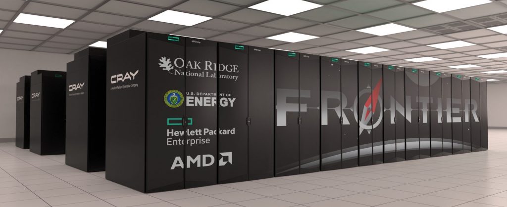
The Infrastructure for Scientific Applications and Advanced Computing (ISAAC) at UT offers both secure and non-secure enclaves containing 1000s of advanced computer cores and multi-GPU nodes integrated with a high-speed Lustre file system with petabyte scale storage capacity. ISAAC is connected to the larger US academic internet through Internet2 connectivity. We also use the OLCF supercomputing resources at ORNL which include the exascale Frontier supercomputer, the current World number 1, as well as other national facilities.
Principal invesitgators: Profs. Dagotto, Del Maestro, and Johnston